9 0 5 7 8
论文已发表
注册即可获取德孚的最新动态
IF 收录期刊
- 2.6 Breast Cancer (Dove Med Press)
- 3.9 Clin Epidemiol
- 3.3 Cancer Manag Res
- 3.9 Infect Drug Resist
- 3.6 Clin Interv Aging
- 4.8 Drug Des Dev Ther
- 2.8 Int J Chronic Obstr
- 8.0 Int J Nanomed
- 2.3 Int J Women's Health
- 3.2 Neuropsych Dis Treat
- 4.0 OncoTargets Ther
- 2.2 Patient Prefer Adher
- 2.8 Ther Clin Risk Manag
- 2.7 J Pain Res
- 3.3 Diabet Metab Synd Ob
- 4.3 Psychol Res Behav Ma
- 3.4 Nat Sci Sleep
- 1.9 Pharmgenomics Pers Med
- 3.5 Risk Manag Healthc Policy
- 4.5 J Inflamm Res
- 2.3 Int J Gen Med
- 4.1 J Hepatocell Carcinoma
- 3.2 J Asthma Allergy
- 2.3 Clin Cosmet Investig Dermatol
- 3.3 J Multidiscip Healthc

长期缺乏长时间睡眠降低健康男性受试者的默认自发活动和连接方式: 静息态功能磁共振成像(fMRI)研究
Authors Dai XJ, Liu CL, Zhou RL, Gong HH, Wu B, Gao L, Wang YX
Published Date March 2015 Volume 2015:11 Pages 761—772
DOI http://dx.doi.org/10.2147/NDT.S78335
Received 28 November 2014, Accepted 3 February 2015, Published 19 March 2015
Objective: The aim of this study is to use resting-state
functional connectivity (rsFC) and amplitude of low-frequency fluctuation
(ALFF) methods to explore intrinsic default-mode network (DMN) impairment after
sleep deprivation (SD) and its relationships with clinical features.
Methods: Twelve healthy male
subjects underwent resting-state functional magnetic resonance imaging twice:
once following rested wakefulness (RW) and the other following 72 hours of
total SD. Before the scans, all subjects underwent the attention network test
(ANT). The independent component analysis (ICA), rsFC, and ALFF methods were
used to examine intrinsic DMN impairment. Receiver operating characteristic
(ROC) curve was used to distinguish SD status from RW status.
Results: Compared with RW
subjects, SD subjects showed a lower accuracy rate (RW =96.83%, SD =77.67%; P <0.001), a slower reaction time
(RW =695.92 ms; SD =799.18 ms; P =0.003), a higher lapse rate (RW =0.69%, SD =19.29%; P <0.001), and
a higher intraindividual coefficient of variability in reaction time (RW =0.26,
SD =0.33; P =0.021). The ICA method showed
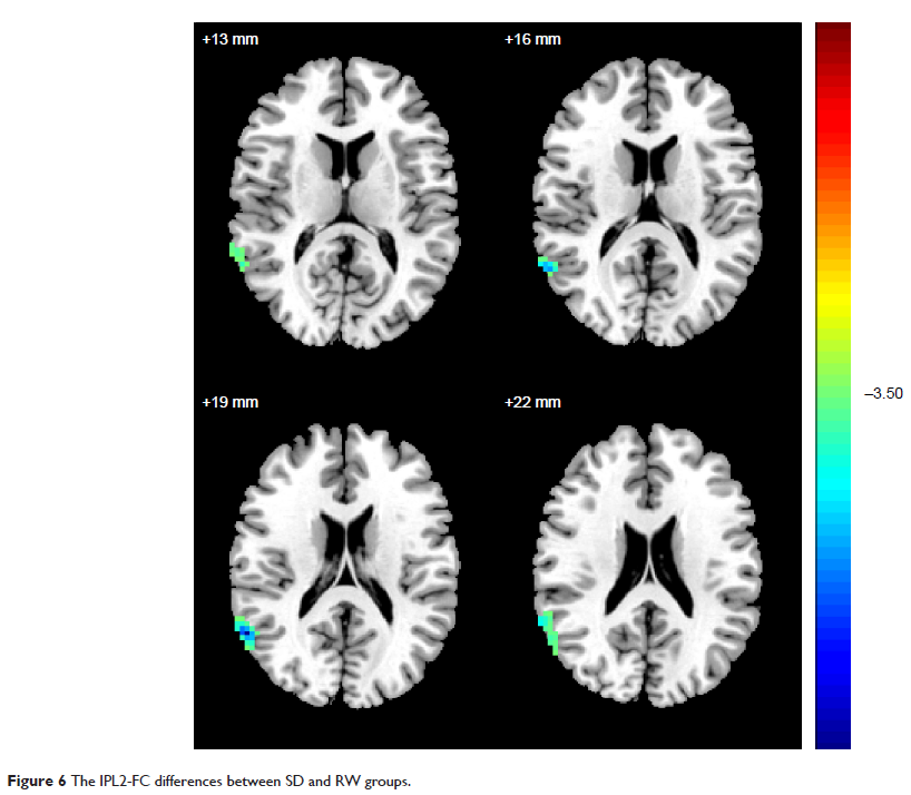
that, compared with RW subjects, SD subjects had decreased rsFC in the right
inferior parietal lobule (IPL, BA40) and in the left precuneus (PrC)/posterior
cingulate cortex (PCC) (BA30, 31). The two different areas were selected as
regions of interest (ROIs) for future rsFC analysis. Compared with the same in
RW subjects, in SD subjects, the right IPL showed decreased rsFC with the left
PrC (BA7) and increased rsFC with the left fusiform gyrus (BA37) and the left
cluster of middle temporal gyrus and inferior temporal gyrus (BA37). However, the
left PrC/PCC did not show any connectivity differences. Compared with RW
subjects, SD subjects showed lower ALFF area in the left IPL (BA39, 40). The
left IPL, as an ROI, showed decreased rsFC with the right cluster of IPL and
superior temporal gyrus (BA39, 40). ROC curve analysis showed that the area
under the curve (AUC) value of the left IPL was 0.75, with a cutoff point of
0.834 (mean ALFF signal value). Further diagnostic analysis exhibited that the
AUC alone discriminated SD status from RW status, with 75% sensitivity and
91.7% specificity.
Conclusion: Long-term SD
disturbed the spontaneous activity and connectivity pattern of DMN.
Keywords: sleep deprivation,
amplitude of low-frequency fluctuation, default-mode network, functional
magnetic resonance imaging, functional connectivity, independent component
analysis, receiver operating characteristic curve